Researchers at the University of Wisconsin-Madison in Madison, Wis., said they are proud to publish a groundbreaking paper on a new MRI machine learning network.
They determined how bright color scans can help surgeons detect and accurately remove intracerebral hemorrhage (ICH), or bleeding in the brain.
Walter Block, professor of medical physics and biomedical engineering, led a research team that developed a special algorithm to support doctors who must operate quickly and accurately to extract brain hemorrhage.
The trick is to visualize it and quantify it so the surgeon has the information he needs, Block said.
Tom Lilleholm, PhD candidate and lead author of the research, developed a specific algorithm for the new color-coded MRI machine learning network.
“We got very high accuracy segmentations from the machine here, 96% accurate clot, 81% accurate edema,” he said, showing one of the MRI slides.
Lilieholm said he can show the surgeon how much bleeding he can safely remove in less than a minute.
It’s very helpful to have that and to have robust data for comparison, Liliholm said. That’s where Matt kind of came in.
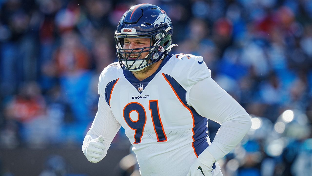
Matt Lilleholm, who he had in mind, was NFL player Matt Henningsen.
Henningsen is from Menomonee Falls. Before becoming a Denver Broncos, he attended UW-Madison, where he excelled on the football field and in the classroom. He received his bachelor’s and master’s degrees from the university.
“My job is to identify the location of the intracerebral hemorrhage and divide the clot and the edema around the clot, and then go into the individual layers of that image,” Henningsen said.
Henningsen spent more than 100 hours collecting data for this new brain hemorrhage study. He said he is excited and grateful for the opportunity to be a part of this collaboration.
The bioengineer and UW-trained football player said he hopes the project can eventually support and improve what his football career has feared: traumatic brain injury.
He said that currently concussions cannot be diagnosed with an MRI. But I mean, maybe in the future, if you can, you can use machine learning to potentially detect certain anomalies that the human eye can’t necessarily detect or things like that. Maybe we can get somewhere.
#scientists #NFL #player #develop #MRI #machine #learning #network
Image Source : spectrumnews1.com

